
What is Volume EM?
Volume electron microscopy or volume EM (vEM) refers to a group of recently developed imaging approaches that use scanning and transmission electron microscopy (SEM and TEM) to allow the interrogation of cell and tissue ultrastructure in 3D, at μm to mm volume scales and nm resolutions.
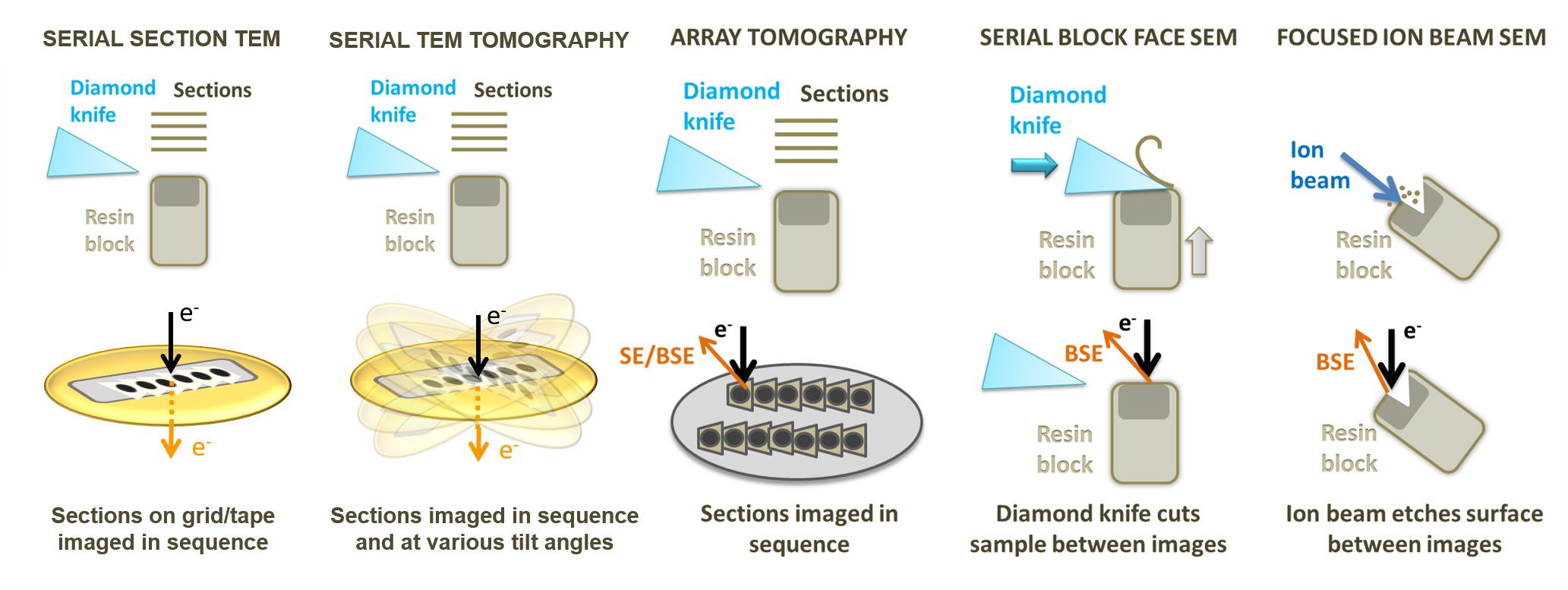
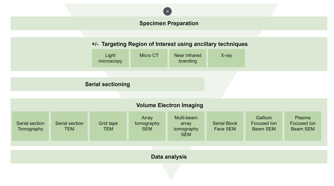
vEM workflows encompass the use of a variety of ancillary techniques both during the preparatory stages and identifying regions of interest, prior to the final electron imaging. Whilst the electron imaging approaches may vary, all vEM workflows share common challenges relating to sample preparation, region of interest targeting and data analysis.
This vEM Community Initiative’s aim is to share knowledge, experience and resources to improve access, reliability, throughput and training of vEM techniques and methodologies.