Serial Block-Face Scanning Electron Microscopy (SBF-SEM or SBEM) focused interest group updates
Organisers: Flavia Moreira-Leite
Organisers: Flavia Moreira-Leite
|
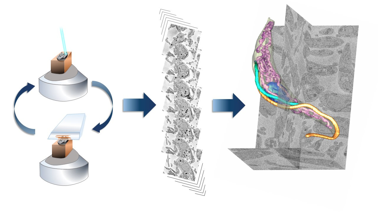
The volume EM technique of SBF-SEM or SBEM allows 3D imaging of relatively large areas of tissue with electron microscopy resolution. In this technique, a resin block containing sample is placed inside a special SEM chamber designed for ultramicrotomy of the block face inside the microscope. In this chamber, the block face can undergo hundreds to thousands of cycles of imaging and sectioning, to generate a large series high resolution SEM images of the same area. These images are then assembled into an aligned 3D volume, which can be used for the 3D reconstruction of whole cells and organelles.
|
With this Focus Interest Group (FIG) we would like to bring together all those interested in SBF-SEM, to discuss, share knowledge and expertise, optimize workflows, build on the advantages of the SBF-SEM techniques, and explore novel fields of application.
We welcome interested parties from academia, research, core facilities and industry.
We welcome interested parties from academia, research, core facilities and industry.

